Image Credits
Illustration And Photo Credits.
Photographs of Dr. Bari and the patient spiral-drawing footage are used with permission. Device images are provided by their manufacturers. The illustrations and photographs below are used under open licenses; each is credited to its creator with a link to the source and license.
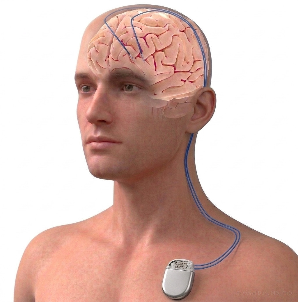 The DBS SystemIllustration supplied by Dr. Bari.
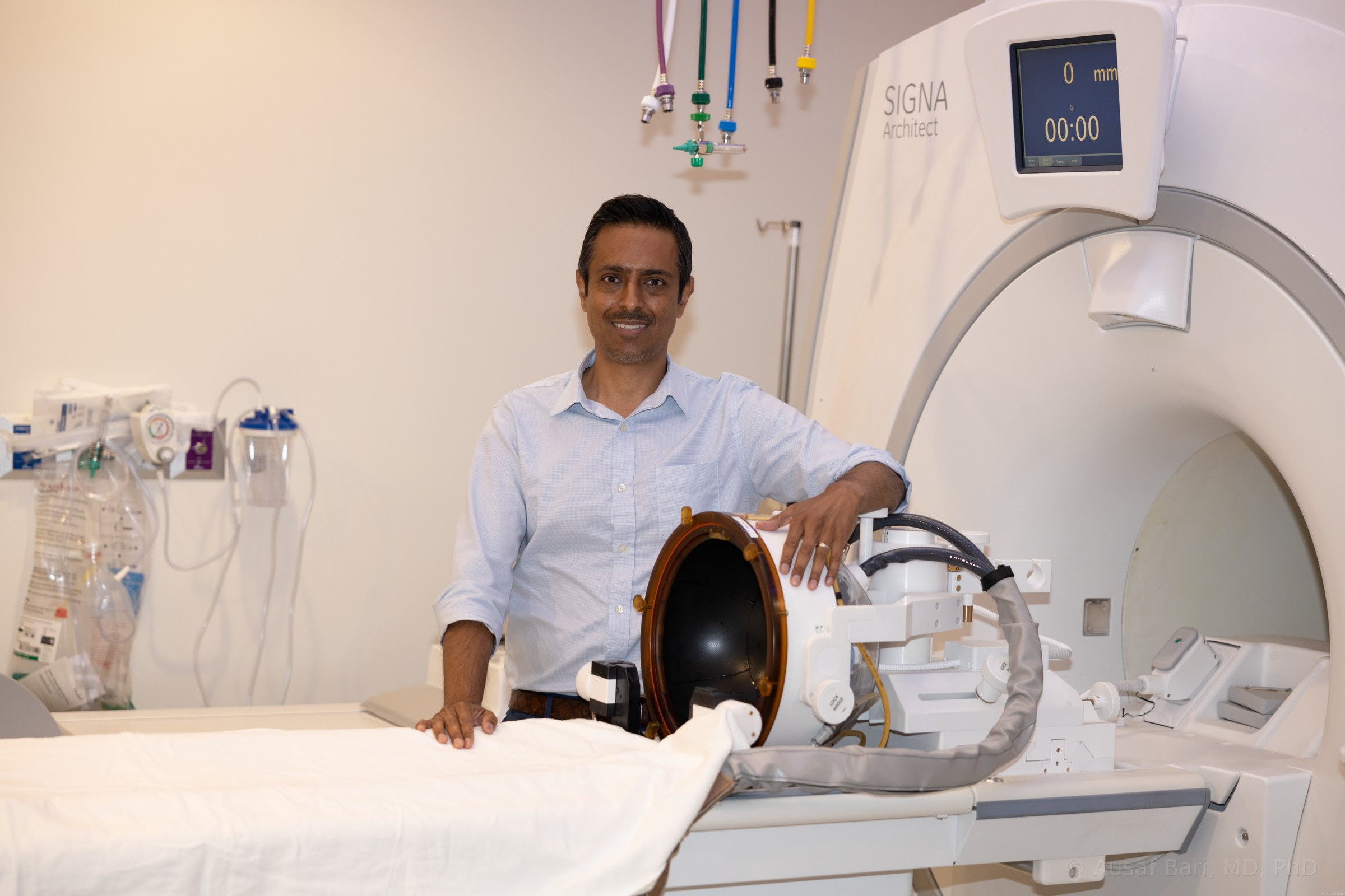 Dr. Bari With The Focused Ultrasound System On The 3T MRIPhotograph from Dr. Bari’s practice.
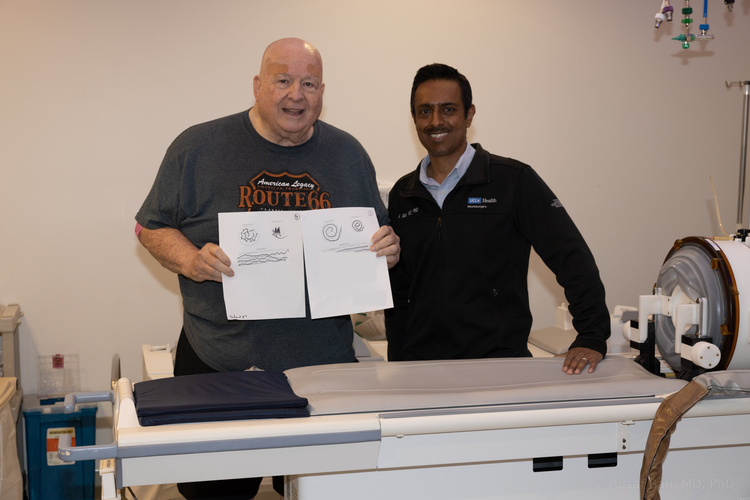 Right After Treatment: Spirals Before (Left) And After (Right)Photograph from Dr. Bari’s practice, used with the patient’s permission. Identifying details removed.
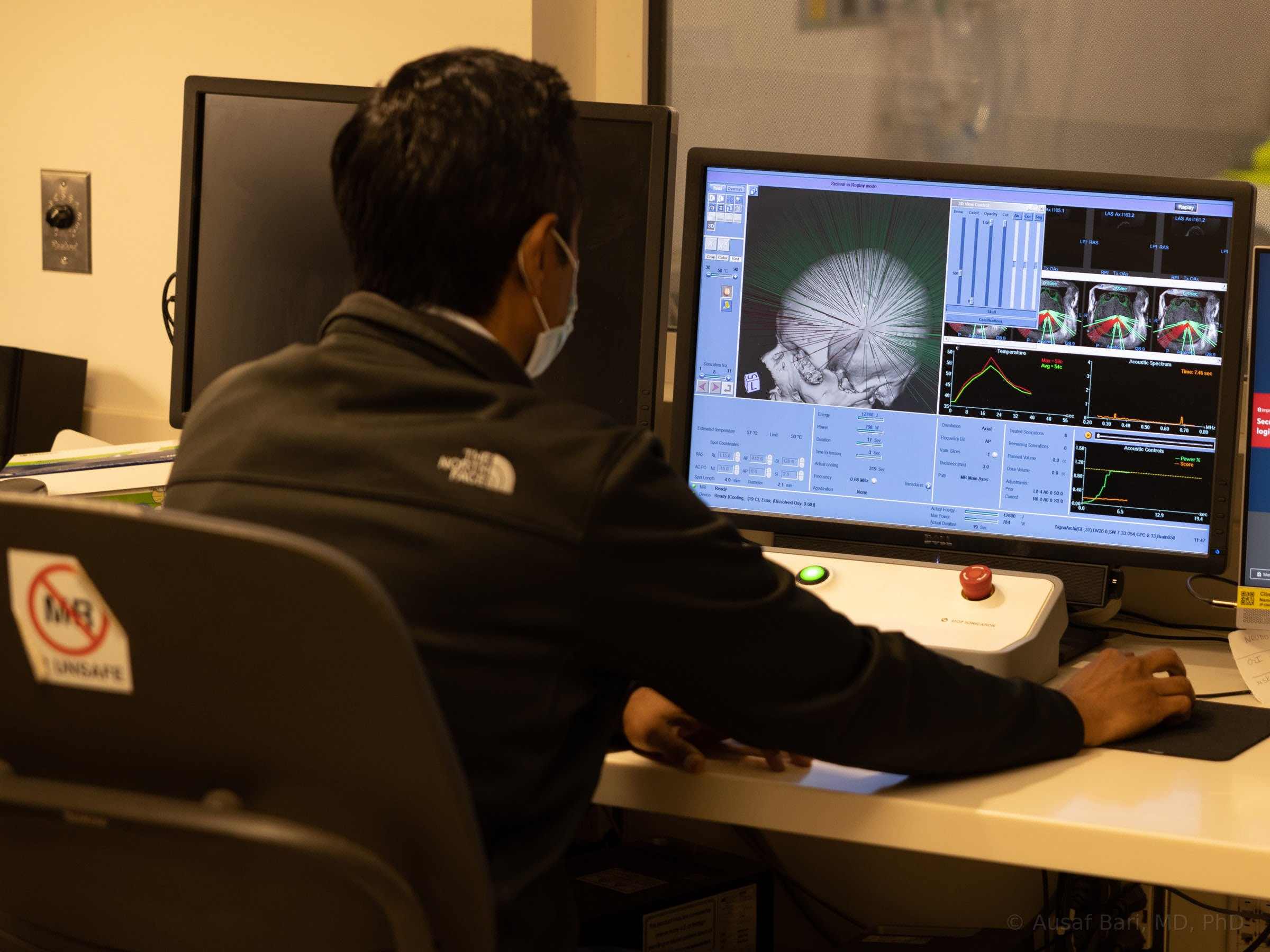 Dr. Bari At The Controls During A Focused Ultrasound TreatmentPhotograph from Dr. Bari’s practice.
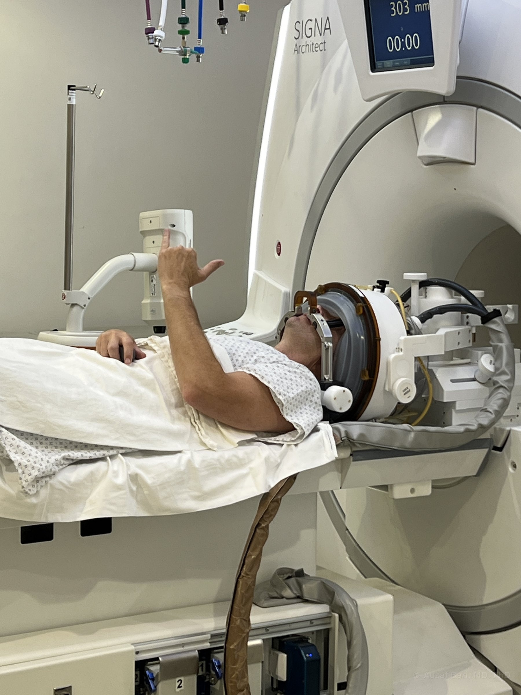 In The Helmet: Awake And Able To Respond During TreatmentPhotograph from Dr. Bari’s practice, used with the patient’s permission.
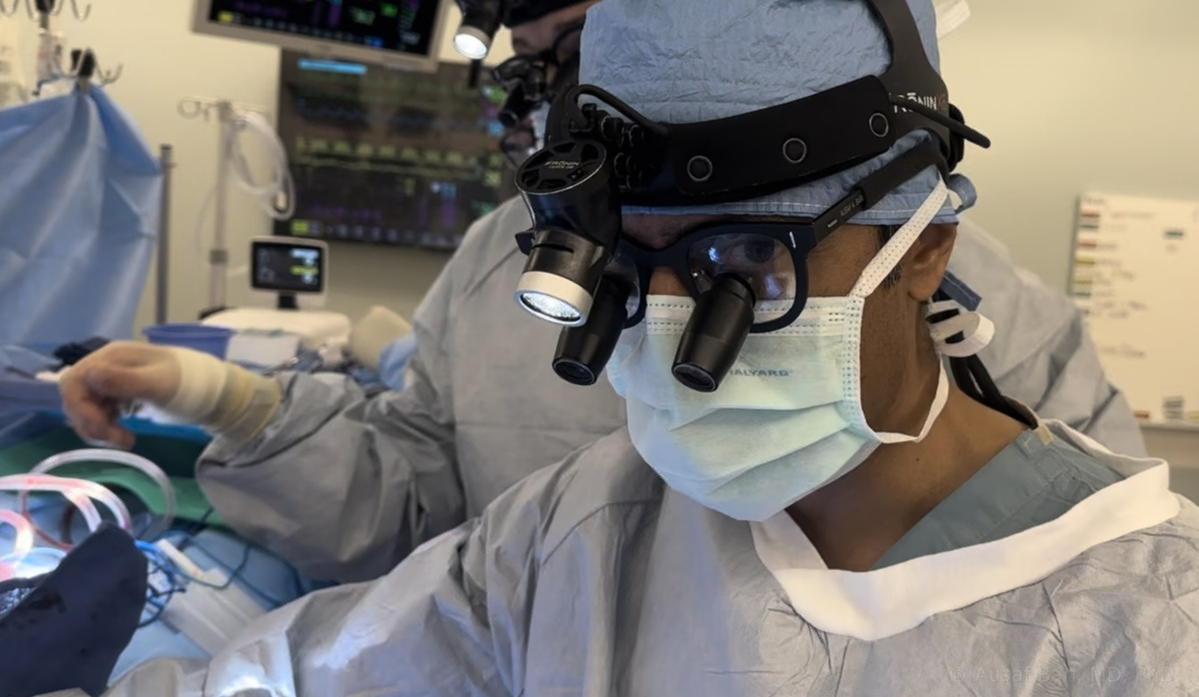 Dr. Bari In The Operating RoomPhotograph from Dr. Bari’s practice.
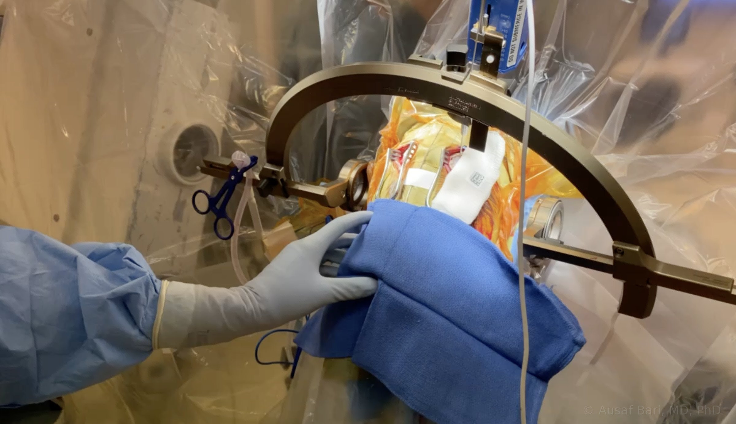 During Deep Brain Stimulation SurgeryPhotograph from Dr. Bari’s practice.
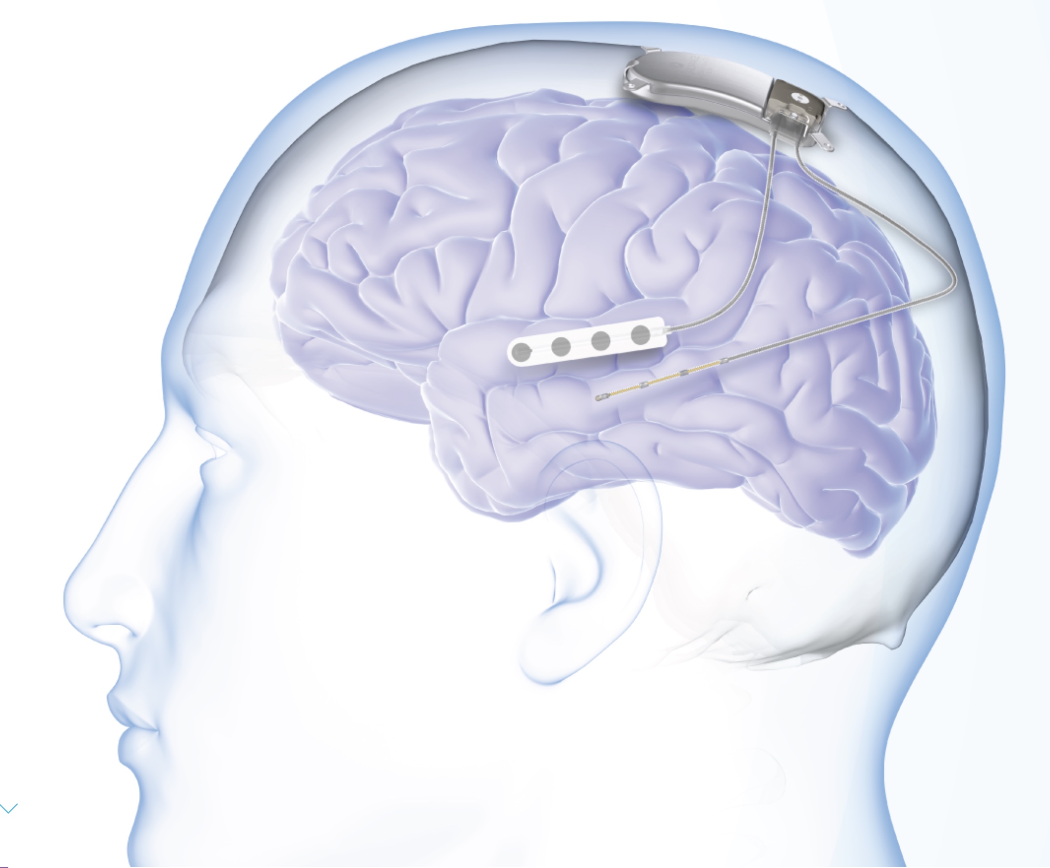 Responsive Neurostimulation For EpilepsyNeuroPace RNS System device image.
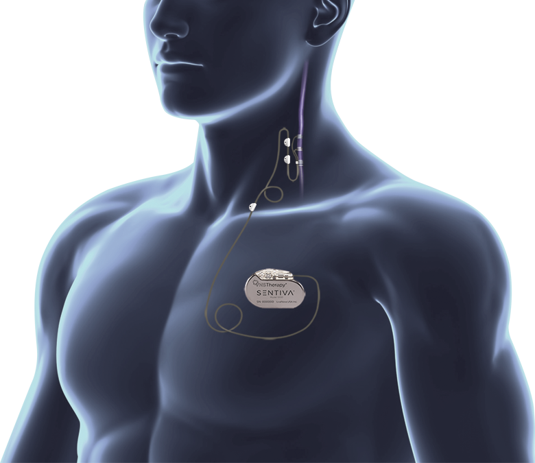 VNS Therapy For Epilepsy: Generator And LeadLivaNova VNS Therapy (SenTiva) device image.
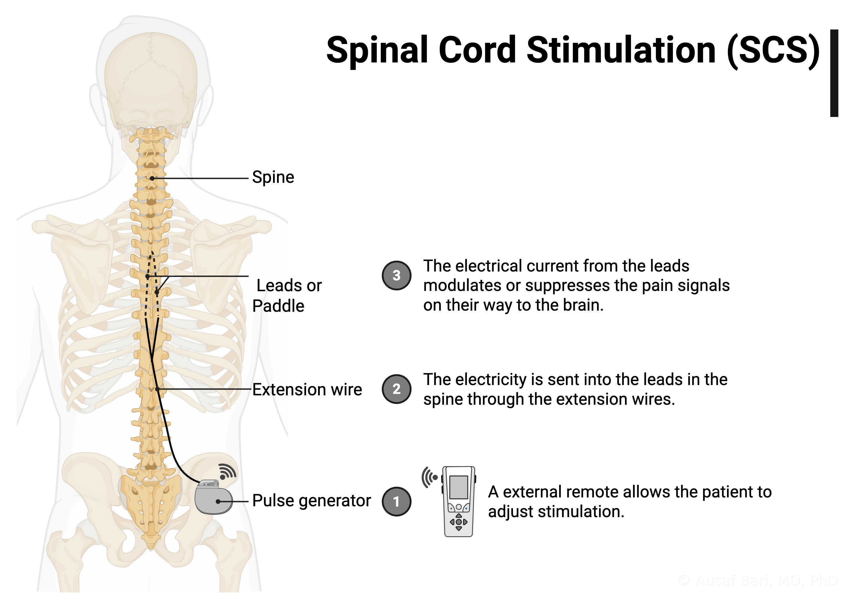 How A Spinal Cord Stimulation System Fits TogetherIllustration supplied by Dr. Bari.
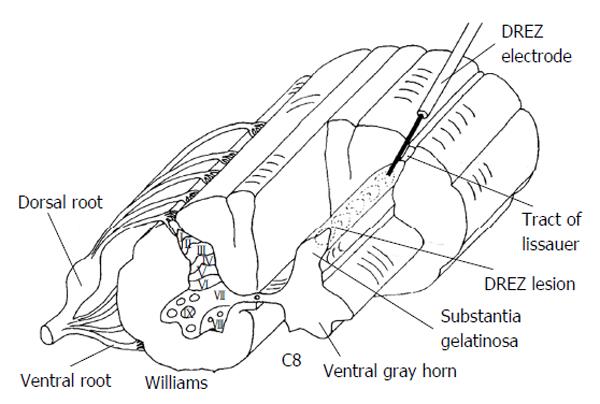 Where The DREZ Lesion Is MadeFrom Dr. Bari’s teaching slides. Figure 4 from Lake and Konrad, World Journal of Surgical Procedures 2015;5(1):111, CC BY-NC 4.0. Unmodified.
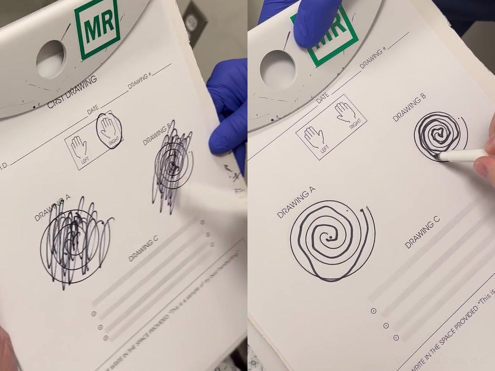 Spiral Drawing: Before (Left), After (Right)Still frames from Dr. Bari’s own clinical footage. One patient example; response varies from patient to patient.
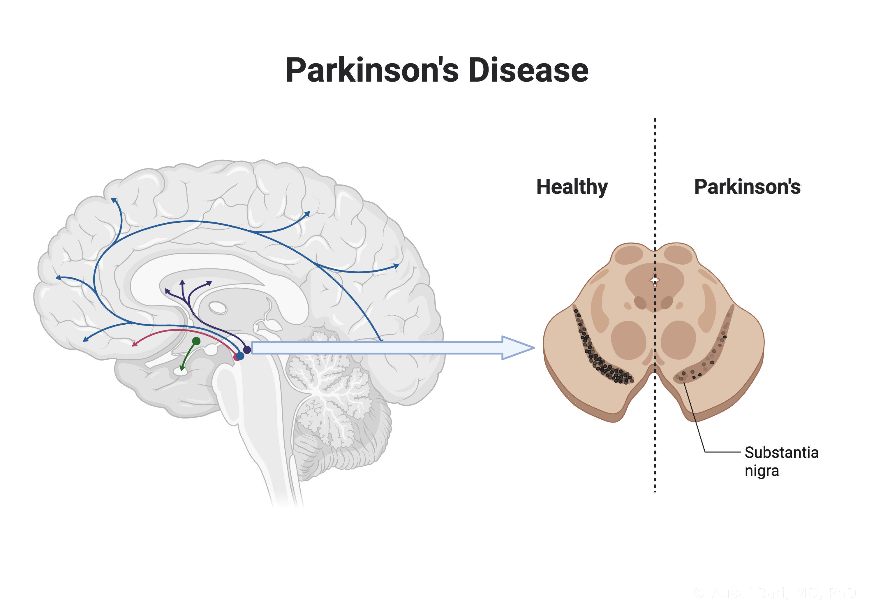 Parkinson’s Disease: Dopamine Pathways And The Substantia NigraIllustration supplied by Dr. Bari.
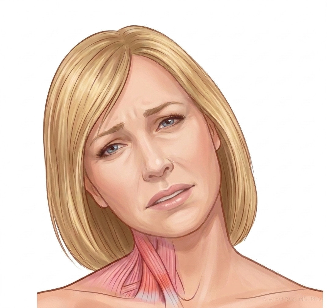 Cervical Dystonia: The Head Pulled To One SideIllustration supplied by Dr. Bari.
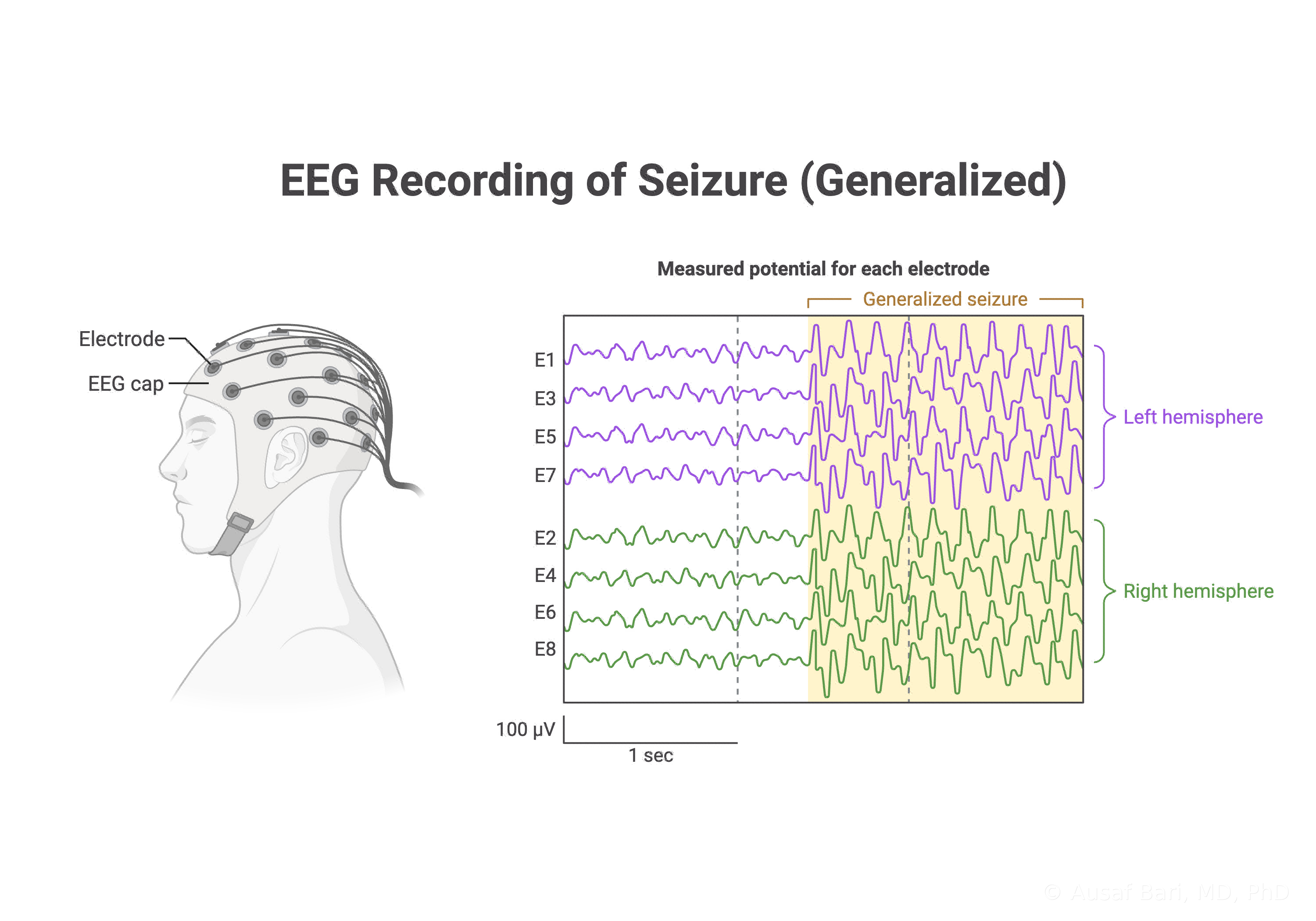 EEG Recording Of A Generalized SeizureIllustration supplied by Dr. Bari.
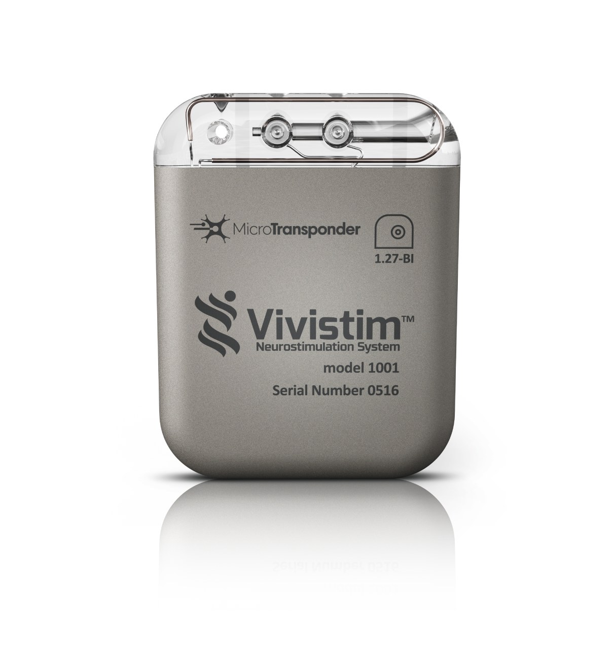 The Vivistim Paired VNS GeneratorMicroTransponder Vivistim device image.
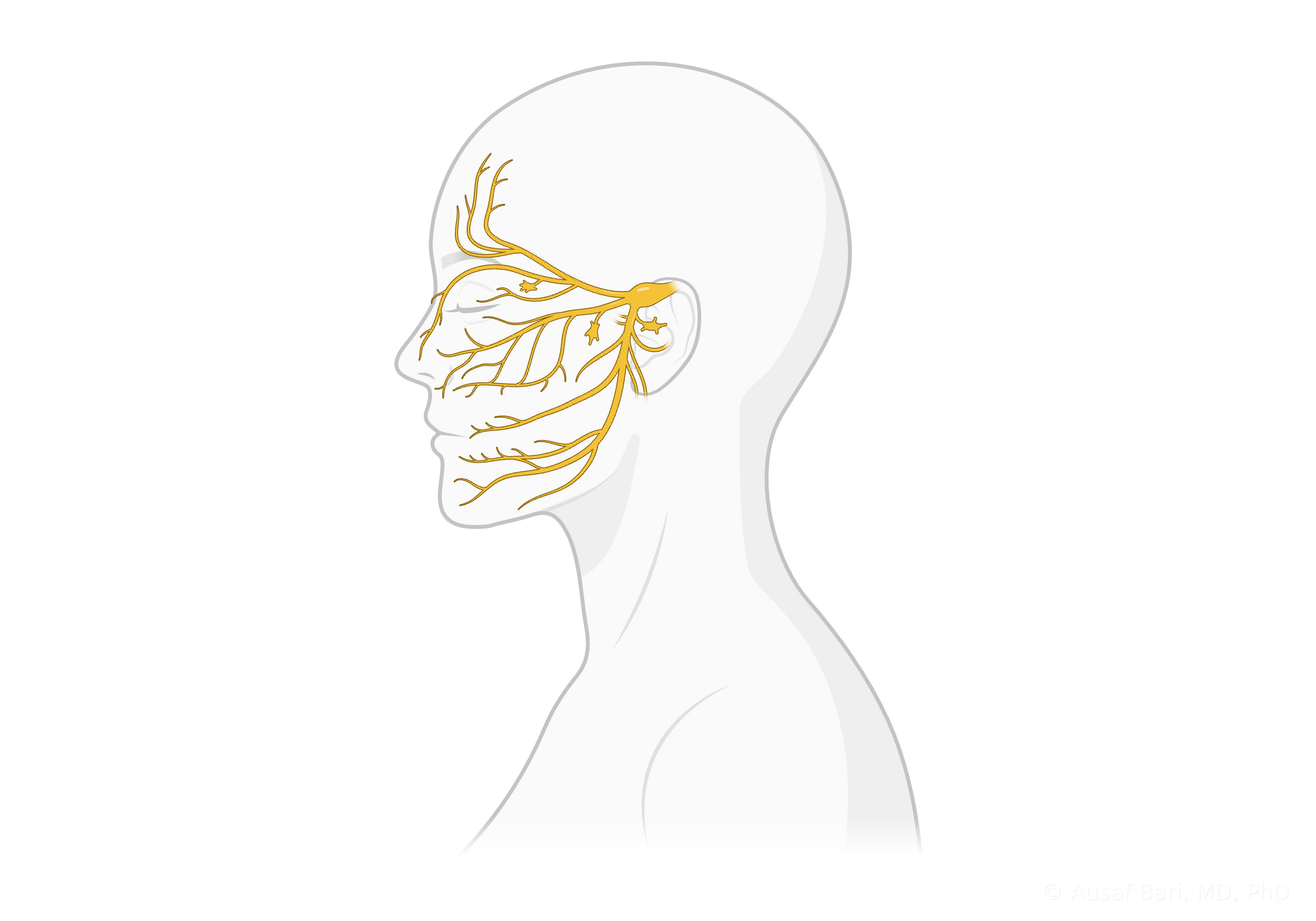 The Trigeminal Nerve And Its BranchesIllustration supplied by Dr. Bari.
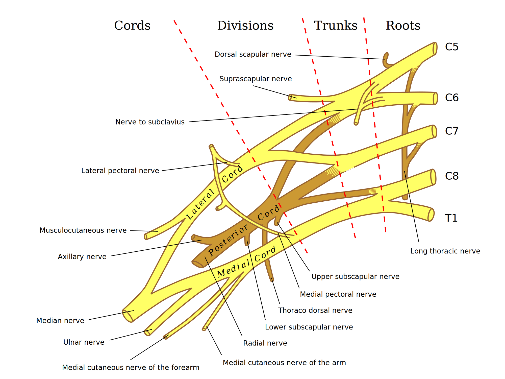 Brachial Plexus AnatomyAfter Gray’s Anatomy (1918); vector by Mattopaedia, Captain-n00dle and MissMJ. Public domain. Rendered from the vector original.
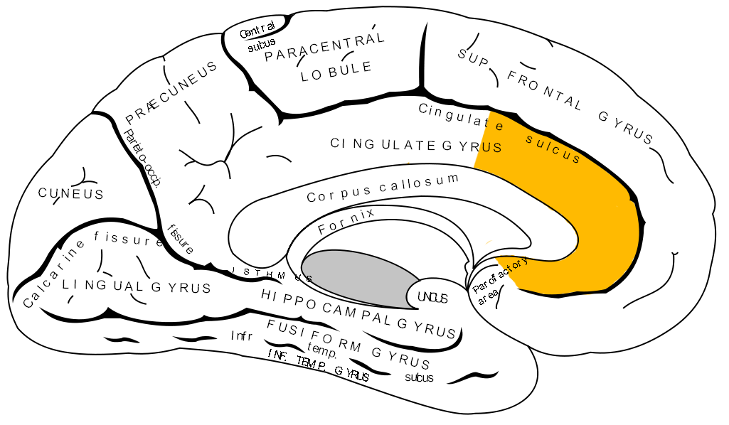 The Anterior Cingulate GyrusGray’s Anatomy, Fig. 727, public domain, via Wikimedia Commons. Unmodified.
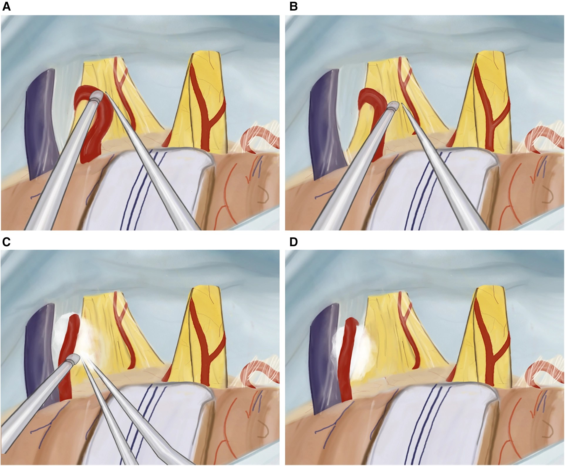 Microvascular Decompression, Step By StepNurimanov et al., Frontiers in Surgery (2024), Figure 4, CC BY 4.0. Unmodified. Panel B shows an additional nerve-combing step that is not part of every operation.
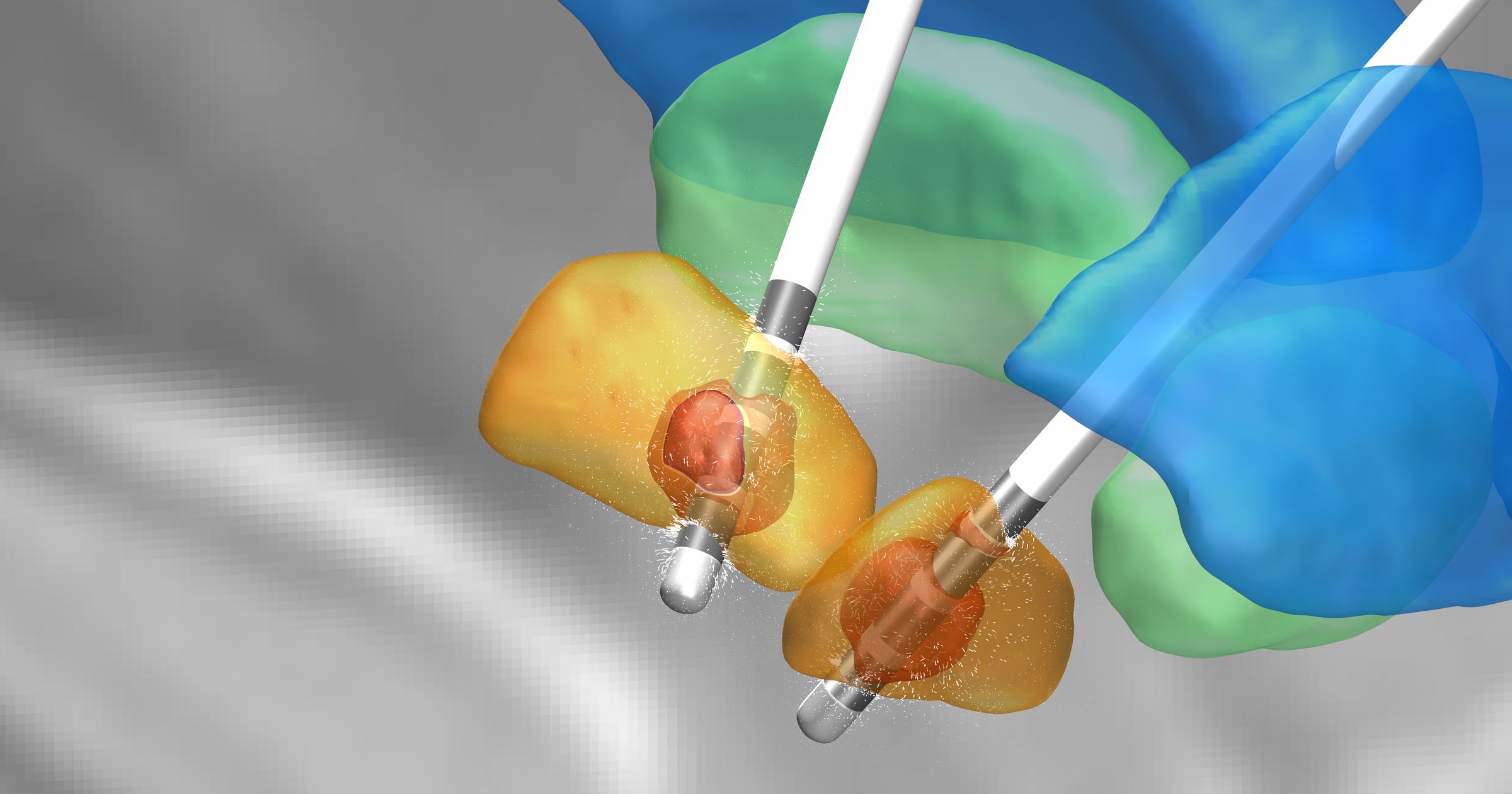 DBS Leads In The Subthalamic NucleusAndreas Horn, CC BY-SA 4.0, via Wikimedia Commons. Resized only.
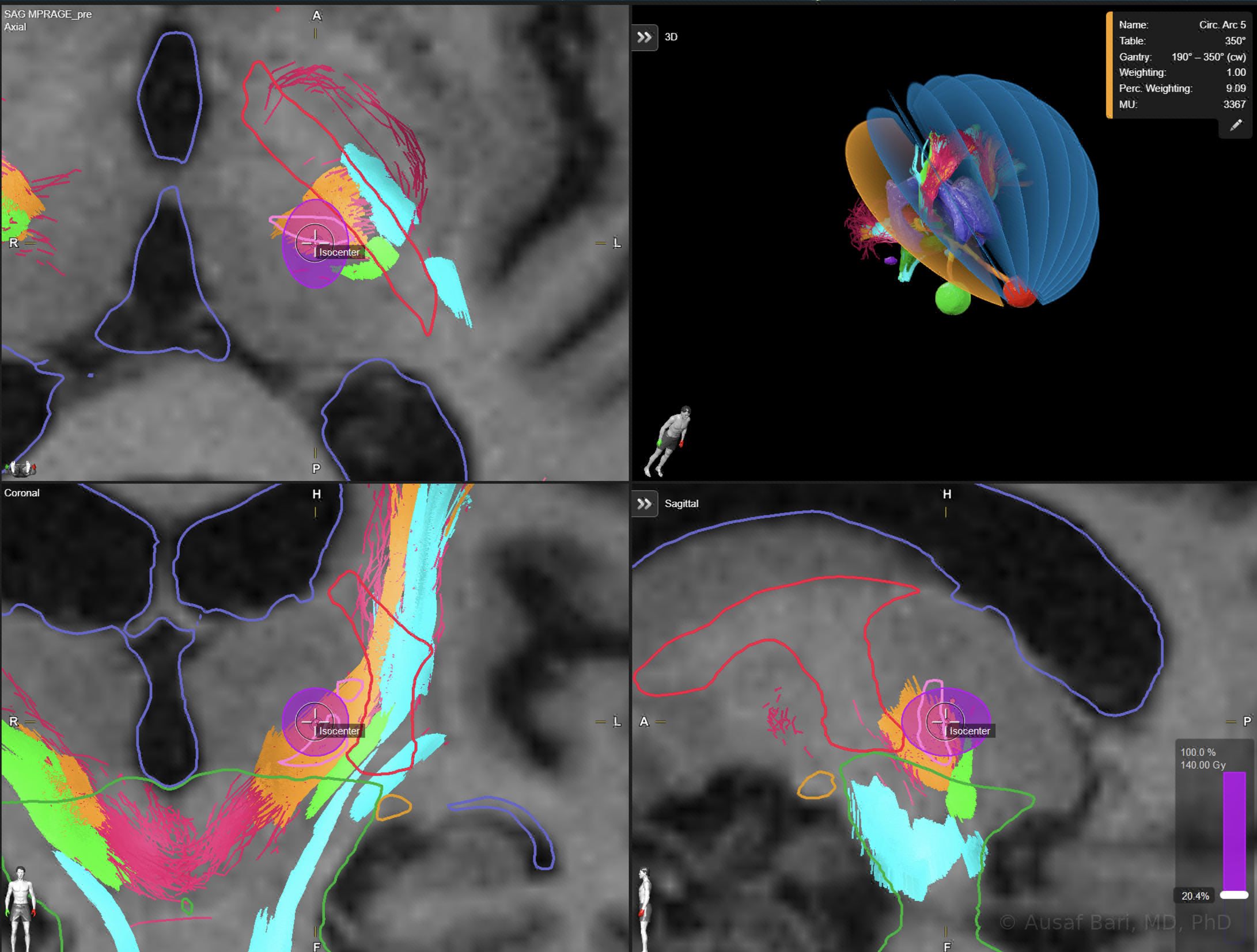 Planning A Radiosurgical Thalamotomy For TremorPlanning image from Dr. Bari’s practice, with no identifying information.
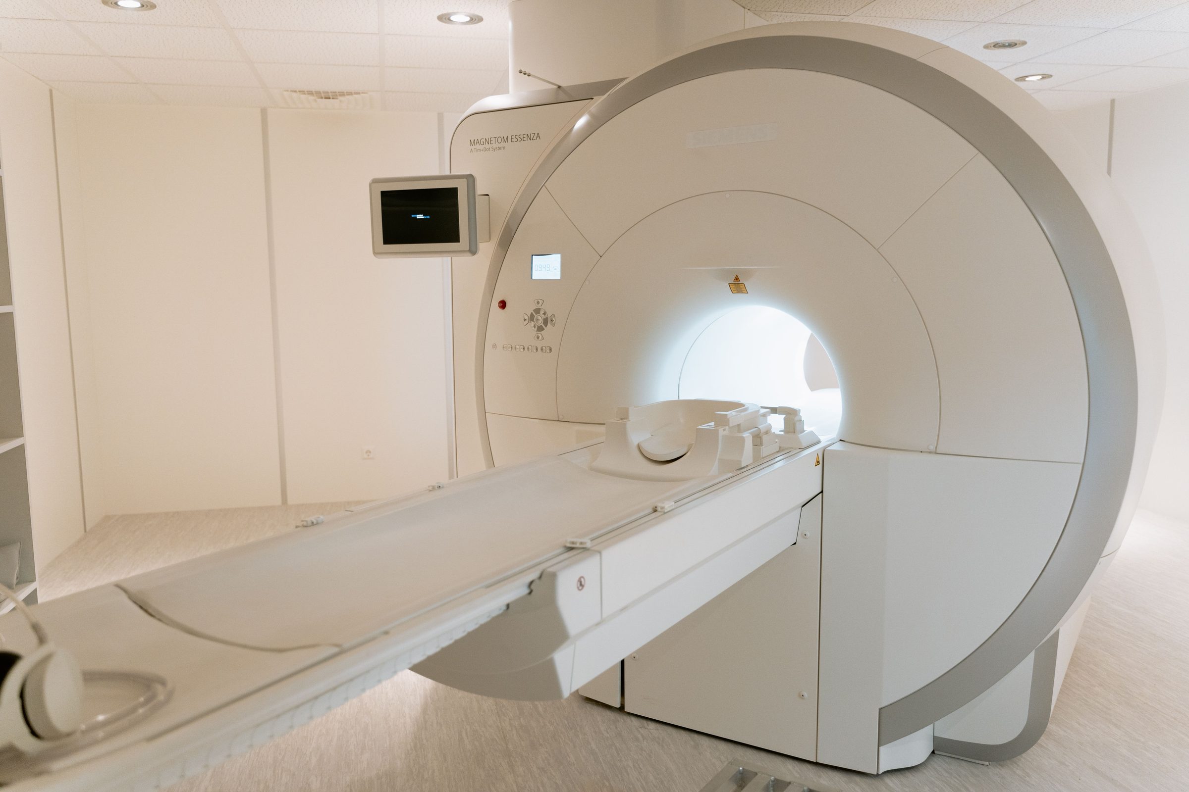 Laser Ablation Is Guided By MRI In Real TimePhoto: MART PRODUCTION, Pexels License.
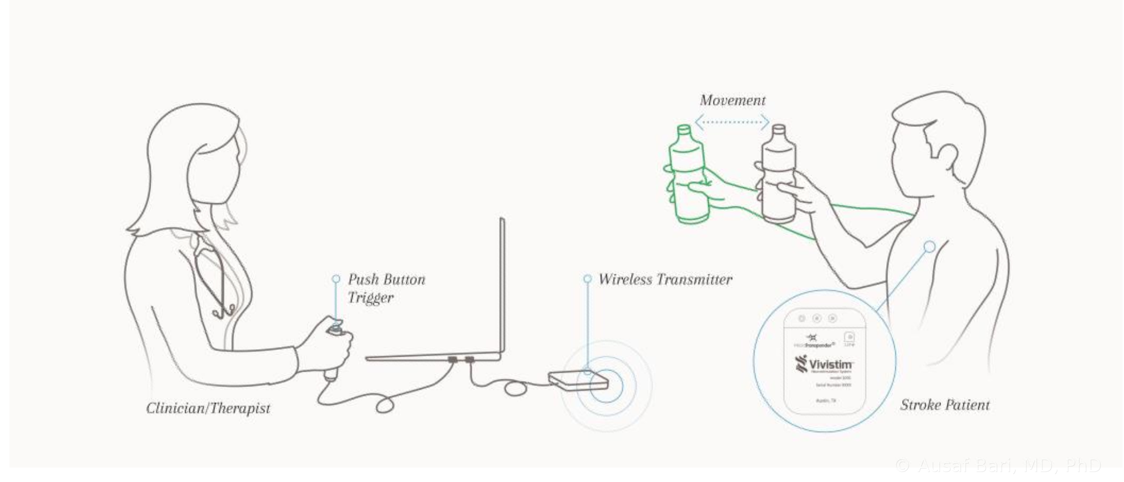 Pairing Stimulation With Arm RehabilitationVivistim System illustration from Dr. Bari’s VNS teaching materials.
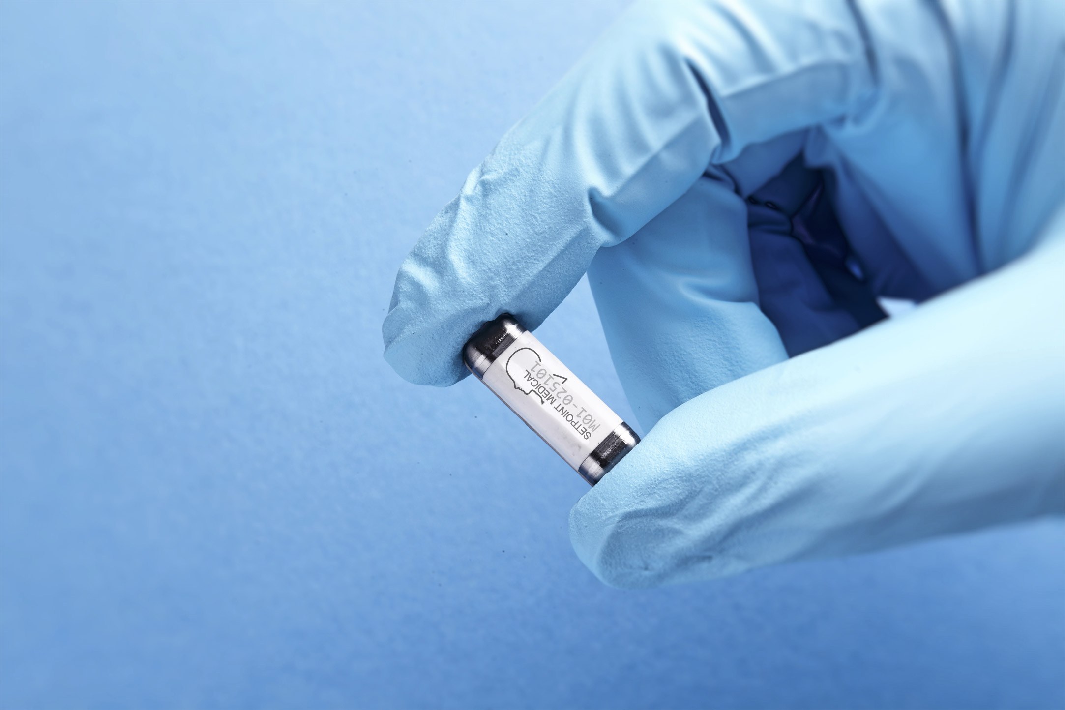 The SetPoint System ImplantSetPoint Medical device image.
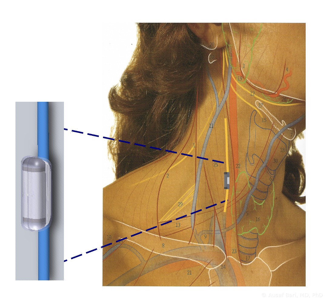 The SetPoint Implant On The Vagus NerveSetPoint Medical illustration from Dr. Bari’s teaching slides.
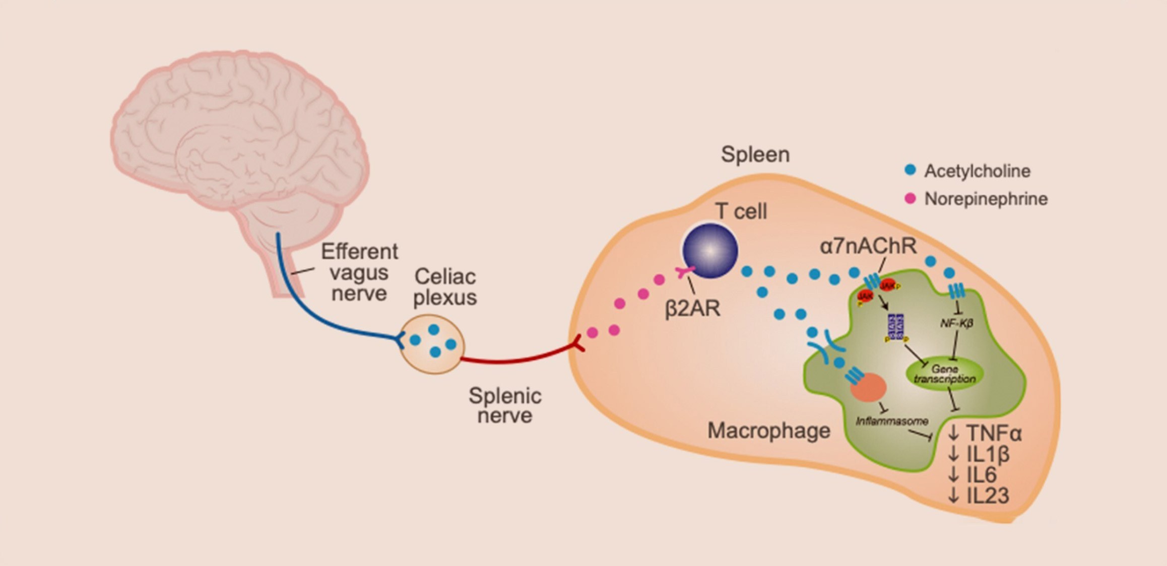 The Inflammatory Reflex: Vagus Nerve To SpleenSetPoint Medical illustration.
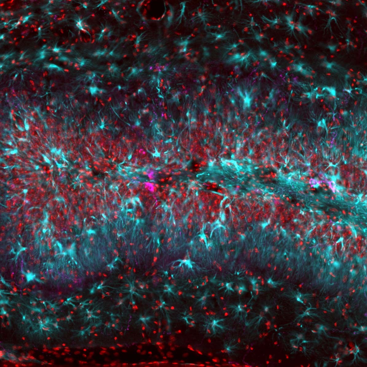 Transplanted Interneurons In The HippocampusNeurona Therapeutics image.
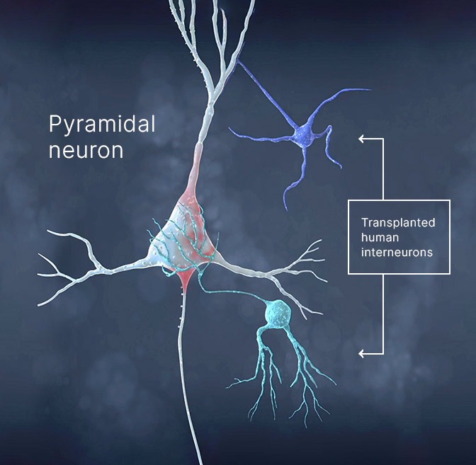 How Transplanted Interneurons Join The CircuitNeurona Therapeutics illustration.
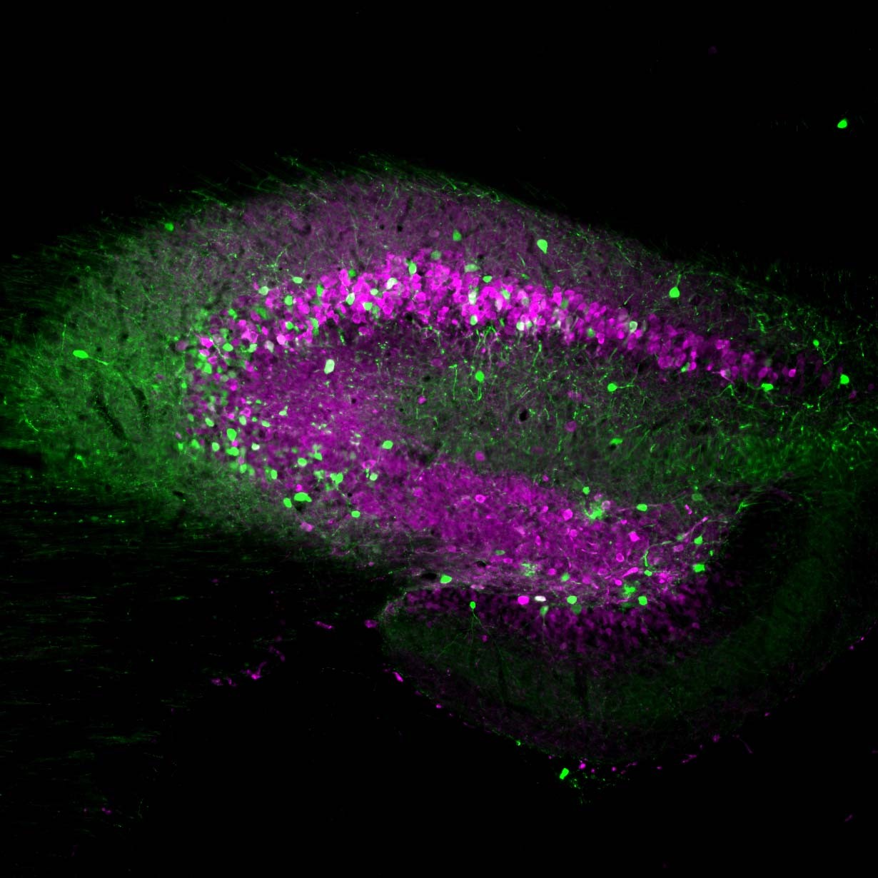 Transplanted Cells Settled Into The Seizure FocusNeurona Therapeutics image.
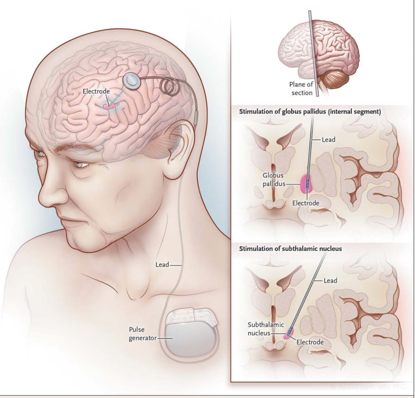 The DBS System And Brain TargetsDevice illustration from Dr. Bari’s DBS teaching materials. The target depends on the condition and individual evaluation.
- An MRI scanner in a quiet, modern imaging suitePhoto: MART PRODUCTION, Pexels License.
 Close-up of an older adult’s hand writing with a penPhoto: RDNE Stock project, Pexels License.
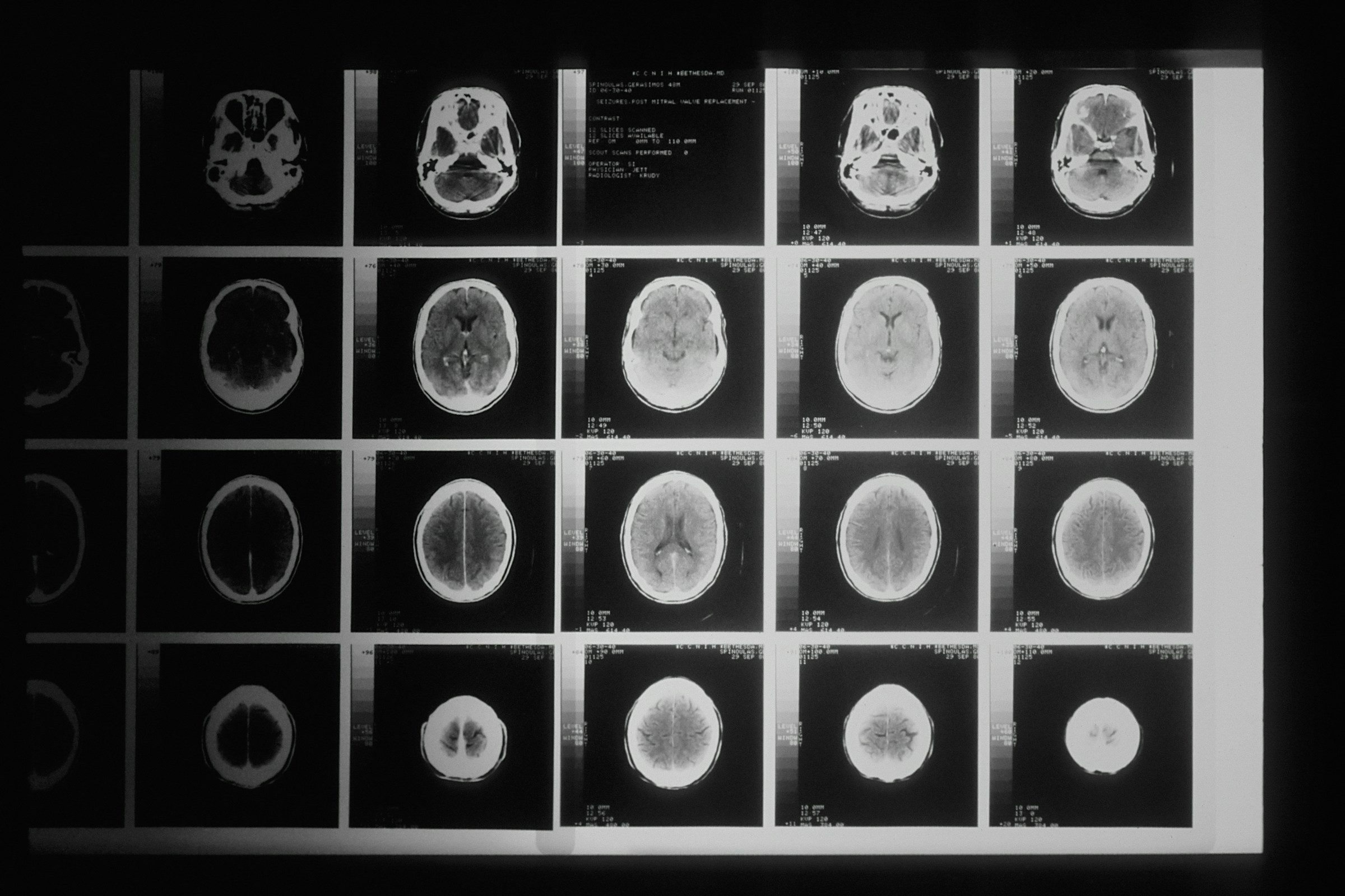 A film of head CT images on a light boxPhoto: National Cancer Institute, Unsplash License.
 The downtown Los Angeles skyline under a hazy blue skyPhoto: Kevin Zhong, Unsplash License.
 Silhouette of a scientist at a microscope against a deep blue backgroundPhoto: National Cancer Institute, Unsplash License.
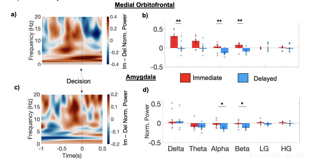 Neural activity during immediate and delayed reward decisionsBrain oscillations recorded around immediate and delayed reward choices in Dr. Bari’s lab.
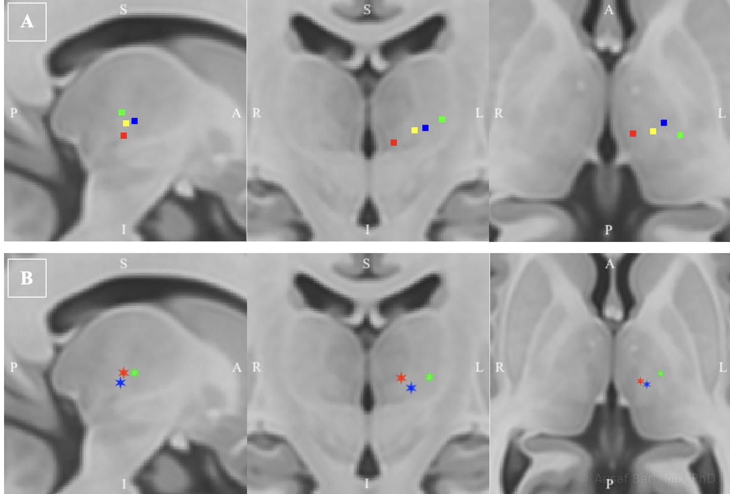 Six brain atlas views with colored markers showing mean thalamic locations associated with body and facial sensory regionsWhere stimulation evoked sensation in different body and facial regions, mapped across patients by Dr. Bari and colleagues (Rifi et al., 2023).
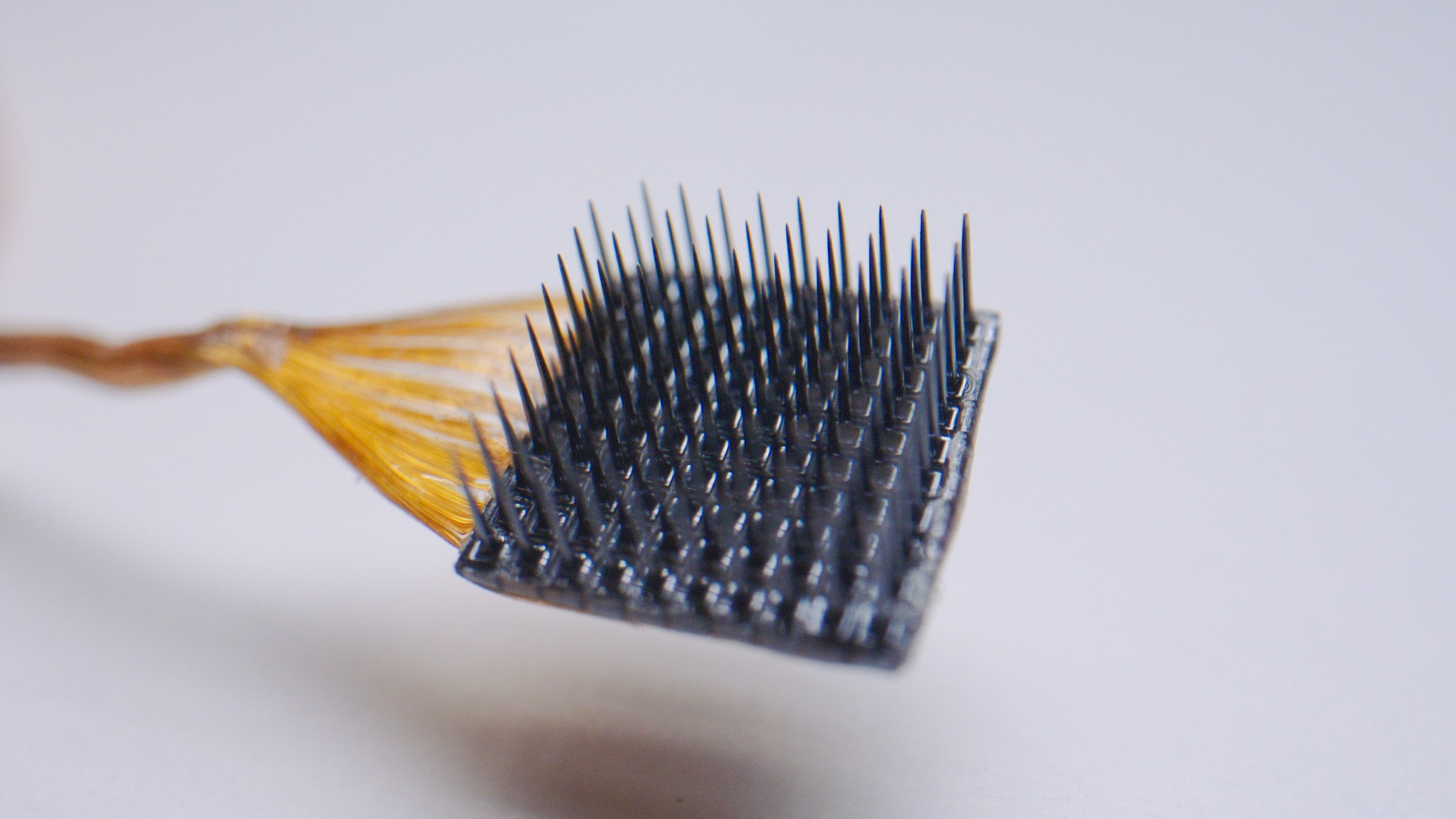 Close-up photograph of a Utah array: a square silicon chip about four millimeters across with a grid of one hundred hair-thin electrode needles and a bundle of gold wiresA Utah array, the intracortical electrode grid Dr. Bari implants in the brain-computer interface trial with Richard Andersen’s lab at Caltech.
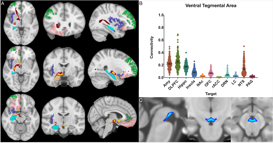 Ventral tegmental area connectivity maps and measured connection strengthsStructural connectivity of the ventral tegmental area across the brain (Levinson et al., 2023).
- Ventral tegmental area connectivity mapsLevinson et al., Frontiers in Neuroimaging (2023), Figure 7, Bari co-author. CC BY 4.0. Unmodified; 2024 correction applied to the caption.


